Valley Fever, also known as coccidioidomycosis, is an emerging concern for veterinarians in the United States, particularly in regions with arid climates such as California.
Current Status of Valley Fever in Dogs
Valley Fever is caused by the soil-dwelling fungi Coccidioides immitis and Coccidioides posadasii. These pathogens are endemic to the southwestern United States, including:
- California
- Arizona
- Nevada
- New Mexico
- Texas
- and Utah
The incidence of Valley Fever in dogs has been rising, paralleling the increase in human cases. Factors contributing to this trend include population growth in endemic areas, increased recreational activities in desert regions, and climate change influencing the distribution and virulence of the fungi.
In California, the Central Valley and surrounding regions are hotspots for Valley Fever. The disease poses a significant health risk to dogs living in or traveling through these areas. Recent studies indicate that approximately 30-40% of dogs residing in endemic regions are exposed to the fungi at some point in their lives, with a subset developing clinical disease.
Clinical Presentation of Valley Fever in Dogs
Dogs infected with Valley Fever can present with a variety of clinical signs, which can be acute or chronic. The severity and type of symptoms depend on the extent of fungal dissemination and the organs affected. Here are the common clinical presentations in canine patients:
- Respiratory Symptoms: Coughing, labored breathing, nasal discharge, and lethargy.
- Musculoskeletal Symptoms: Lameness, swelling of limbs, bone pain, and reluctance to move.
- Cutaneous Symptoms: Skin lesions, draining tracts, and non-healing sores.
- Neurological Symptoms: Seizures, ataxia, and changes in behavior or consciousness.
- General Symptoms: Fever, weight loss, anorexia, and generalized weakness.
It is important for veterinarians to consider Valley Fever in the differential diagnosis when dogs from endemic areas present with these non-specific signs.
Pathogens Causing Valley Fever in Dogs
Valley Fever is caused by two primary pathogens:
- Coccidioides immitis: Predominantly found in California’s San Joaquin Valley and surrounding areas.
- Coccidioides posadasii: More commonly found in other parts of the southwestern United States, including Arizona.
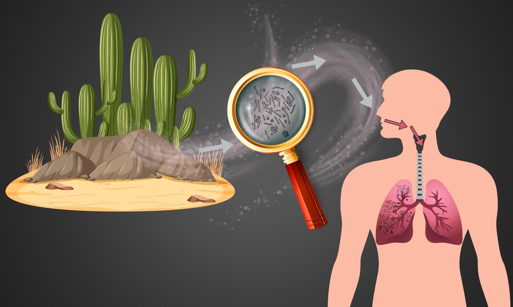
Both species have similar pathogenicity and clinical manifestations in dogs. The fungi exist in the soil as arthroconidia, which are inhaled by the host. Once in the lungs, the arthroconidia transform into spherules that contain endospores, leading to local infection and potential dissemination to other organs.
Diagnostic Tools for Valley Fever
Accurate and timely diagnosis of Valley Fever in dogs is crucial for effective treatment and management. Several diagnostic tools are available to veterinarians:
- Serology: Detection of antibodies against Coccidioides spp. in the blood using tests such as enzyme-linked immunosorbent assay (ELISA), immunodiffusion (ID), and complement fixation (CF). These tests are widely used but can have limitations in sensitivity and specificity.
- Cytology and Histopathology: Examination of tissue samples from affected organs (e.g., lymph nodes, bone, skin) to identify spherules of Coccidioides spp. This method is direct but may require invasive procedures to obtain samples.
- Culture: Isolation of Coccidioides spp. from clinical specimens (e.g., respiratory secretions, tissue biopsies). Culture is definitive but slow and poses a biohazard risk to laboratory personnel.
- Polymerase Chain Reaction (PCR): Detection of fungal DNA in clinical samples. PCR offers rapid results and high specificity but may not always be available in all veterinary laboratories.

Next-Generation Sequencing as a Diagnostic Tool
Next-generation sequencing (NGS) has emerged as a powerful diagnostic tool with higher sensitivity and specificity for detecting Valley Fever compared to traditional methods. NGS allows for the comprehensive analysis of genetic material in clinical samples, enabling the identification of Coccidioides spp. even in low-abundance infections.
Advantages of NGS in Diagnosing Valley Fever
- Higher Sensitivity: NGS can detect Coccidioides DNA at very low levels, increasing the likelihood of diagnosing infections that might be missed by serology or PCR.
- Higher Specificity: By analyzing the genetic material, NGS can accurately distinguish between different pathogens and provide definitive identification of Coccidioides spp.
- Comprehensive Detection: NGS can simultaneously identify multiple pathogens, which is particularly useful in cases where co-infections are suspected.
- Non-Invasive Sampling: NGS can be performed on various sample types, including blood, respiratory secretions, and tissue biopsies, reducing the need for invasive procedures.
Implementation of NGS in Veterinary Practice
The use of NGS in veterinary diagnostics is gaining traction, with several commercial laboratories offering NGS-based testing for Valley Fever and other infectious diseases. Veterinarians should consider the following when implementing NGS:
- Sample Collection and Handling: Proper collection and preservation of samples are crucial for accurate NGS results. By using the MiDOG collection kits, the samples are preserved at room temperature and the pathogens are effectively lysed upon contact for save handling of the sample for all personnel involved.
- Turnaround Time: While NGS provides comprehensive results, with a turnaround time of just 2-3 days by the MiDOG laboratory for requiring rapid diagnosis.
Valley Fever in Dogs: Conclusion
Valley Fever remains a significant health concern for dogs in the USA, particularly in endemic regions like California. Veterinarians play a crucial role in the early detection and management of this disease. Understanding the clinical presentation, causative pathogens, and available diagnostic tools is essential for providing optimal care.
Next-generation sequencing testing, as offered in the All-in-One Test by MiDOG Animal Diagnostics, represents a significant advancement in the diagnosis of Valley Fever, offering higher sensitivity and specificity compared to traditional methods. By incorporating NGS into diagnostic protocols, veterinarians can improve the accuracy of diagnoses and enhance the overall quality of care for their patients.
For further reading and up-to-date information on Valley Fever in dogs, refer to the following sources:
- Shubitz, L.F., & Dial, S.M. (2021). Coccidioidomycosis: A Review of Recent Advances. Journal of Fungi, 7(5), 378. Link
- Thompson, G.R., & Wiederhold, N.P. (2022). Coccidioidomycosis: A Changing Epidemiology. Clinical Microbiology Reviews, 35(3), e00015-22. Link
- Galgiani, J.N., et al. (2020). Valley Fever (Coccidioidomycosis) in Dogs: An Overview for Veterinarians. Veterinary Clinics of North America: Small Animal Practice, 50(2), 341-358. Link
By staying informed and utilizing advanced diagnostic tools, veterinarians can effectively combat Valley Fever and ensure the well-being of their canine patients.