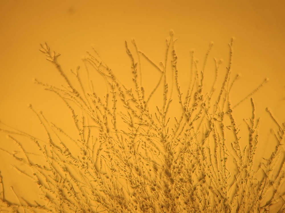
Fusarium species are a group of filamentous fungi widely recognized for their pathogenicity in agriculture, where they primarily infect crops like corn, wheat, and barley. These fungi exhibit typical fungal morphology, characterized by septate hyphae, conidia (asexual spores), and distinctive macro- and microconidia. Fusarium species can produce a variety of toxins, including mycotoxins like fumonisins, trichothecenes, and zearalenone, which have serious implications for both agricultural productivity and animal health. Traditionally, Fusarium was seen as a crop pathogen, but in recent years, its impact has expanded to veterinary medicine. Several species of Fusarium are now known to infect animals, causing a variety of diseases in a broad host range (O’Donnel et al., 2016). Additionally, when Fusarium-contaminated feed is consumed by animals, mycotoxins can contaminate the food chain, leading to toxic effects in livestock and other animals (Placinta et al., 1999). These mycotoxins can lead to immunosuppression, gastrointestinal issues, reproductive disorders, and even death in severe cases. As a result, Fusarium species are increasingly recognized as a significant concern in both food safety and animal health. In this blog post we will explore the veterinary impacts of Fusarium. We discuss the challenges of treating the pathogen and how the fungus’ history as an agricultural pathogen enhances its danger in veterinary disease. Finally, we explain the utility and importance of Next Generation Sequencing (NGS) as a tool for diagnosis of Fusarium.
Major Fusarium Species, Their Animal Hosts, and Symptoms
Several Fusarium species have been identified as causing infections in animals, each linked to particular animal hosts and disease manifestations:
- Fusarium solani
- Hosts: Dogs, cats, horses, cattle, fish
- Symptoms: Keratomycosis (eye infection), mycotic dermatitis, systemic infections (especially in immunocompromised animals)
- Fusarium oxysporum
- Hosts: Cattle, sheep, horses, poultry
- Symptoms: Respiratory issues, gastrointestinal upset, reproductive failure (due to mycotoxin contamination)
- Fusarium verticillioides (formerly Fusarium moniliforme)
- Hosts: Swine, poultry, horses
- Symptoms: Leukoencephalomalacia (brain disease) in horses, growth stunting and neurological symptoms in swine and poultry
- Fusarium graminearum
- Hosts: Cattle, pigs, horses, poultry
- Symptoms: Reproductive failure, anorexia, lethargy, mycotoxin-induced immunosuppression
- Fusarium proliferatum
- Hosts: Swine, poultry
- Symptoms: Liver and kidney damage, gastrointestinal disturbances, reproductive issues
- Fusarium culmorum
- Hosts: Cattle, poultry, swine
- Symptoms: Respiratory disease, digestive problems, mycotoxicosis
Treatment of Fusarium Infections in Animals
When animals are infected by Fusarium species, prompt treatment is essential to mitigate the toxic effects and prevent the spread of the infection. Here are the primary methods of treatment:
- Antifungal Therapy
- Azoles (e.g., itraconazole, fluconazole) are commonly used to treat Fusarium infections. They work by inhibiting the synthesis of ergosterol, a critical component of the fungal cell membrane, thereby disrupting fungal growth (Al-Hatmi et al., 2018).
- Amphotericin B may be administered for more severe, systemic Fusarium infections. This polyene antibiotic binds to ergosterol in the fungal cell membrane, creating pores that lead to cell death (Al-Hatmi et al., 2018).
- Terbinafine is another option, effective against dermatophytes and some Fusarium species, by inhibiting squalene epoxidase, an enzyme involved in the biosynthesis of ergosterol (Al-Hatmi et al., 2018).
- Supportive Care
- Intravenous fluids, nutritional support, and anti-inflammatory drugs (e.g., corticosteroids) help manage symptoms and improve the animal’s recovery chances.
- For cases of systemic infection or intoxication, activated charcoal may be used to limit the absorption of mycotoxins from the gastrointestinal tract.
- Surgical Intervention
- In some cases, surgical debridement or removal of infected tissue, such as abscesses or necrotic tissue caused by Fusarium keratomycosis, may be necessary (Al-Hatmi et al., 2018).
- Detoxification of Animal Feed
- The use of mycotoxin binders (e.g., activated charcoal, clay-based adsorbents) in contaminated animal feed can help reduce the toxic burden on affected animals.
Fungicides to Avoid Due to Fusarium Antifungal Resistance
Fusarium species are notorious for their hardy nature and resistance to several fungicides, a phenomenon that has been exacerbated by certain agricultural practices. The widespread use of fungicides in agriculture has led to the development of resistance in Fusarium, making some treatments ineffective for controlling fungal growth. Here’s a breakdown of the observed antifungal resistance in Fusarium:
- Triazoles (e.g., tebuconazole, propiconazole) – Fusarium species, especially Fusarium solani, have developed resistance mechanisms that render these fungicides ineffective. Overuse of triazoles in agricultural settings, where they are commonly applied to crops like wheat and corn, has selected for Fusarium strains that can metabolize or pump out the fungicide before it can have an effect (Al-Hatmi et al., 2018).
- Strobilurins (e.g., azoxystrobin, pyraclostrobin) – While effective for many fungal pathogens, Fusarium species have shown significant resistance to strobilurins. Agricultural over-reliance on strobilurins for crop protection has contributed to the development of resistant Fusarium strains. These fungicides inhibit mitochondrial respiration in fungi, but Fusarium species have developed alternative metabolic pathways to survive their action.
- Chlorothalonil – Resistance to this broad-spectrum fungicide has been reported in Fusarium, reducing its efficacy in both agricultural and veterinary settings.
- MBC Fungicides (e.g., benomyl, thiophanate-methyl) – These fungicides are often ineffective against Fusarium, as the fungus can quickly develop resistance mechanisms.
The Role of NGS in Fusarium Diagnosis and Treatment
Next Generation Sequencing (NGS) technologies represent a groundbreaking advancement in the diagnosis and treatment of Fusarium infections in animals. NGS allows for comprehensive molecular analysis of Fusarium species and mycotoxins at the genetic level, providing a more precise understanding of the fungal pathogen involved. By using NGS, veterinarians can quickly identify the specific Fusarium species infecting an animal, even distinguishing between pathogenic and non-pathogenic strains. This level of specificity can lead to more targeted treatment strategies, such as the use of antifungal agents that are effective against the identified species. Such accuracy of classification is critical certain species of Fusarium possess resistances that other species lack (Al-Hatmi et al., 2016). Additionally, NGS can detect mycotoxin profiles in animal tissues and feed, helping to determine the exact toxins present and their potential health impacts. This diagnostic tool can also be used to monitor resistance patterns in Fusarium species, guiding treatment plans and enabling veterinarians to avoid ineffective fungicides. In summary, NGS has the potential to revolutionize the management of Fusarium-related diseases in animals, improving both treatment outcomes and preventative measures.
References
- Al-Hatmi, Abdullah MS, Jacques F. Meis, and G. Sybren de Hoog. “Fusarium: molecular diversity and intrinsic drug resistance.” PLoS pathogens4 (2016): e1005464. DOI: https://doi.org/10.1371/journal.ppat.1005464
- Al-Hatmi, A. M., Bonifaz, A., Ranque, S., De Hoog, G. S., Verweij, P. E., & Meis, J. F. (2018). Current antifungal treatment of fusariosis. International Journal of Antimicrobial Agents, 51(3), 326-332. DOI: https://doi.org/10.1016/j.ijantimicag.2017.06.017
- O’Donnell, Kerry, et al. “Veterinary fusarioses within the United States.” Journal of Clinical Microbiology11 (2016): 2813-2819. DOI: https://doi.org/10.1128/jcm.01607-16
- Placinta, C. M., D’Mello, J. F., & Macdonald, A. M. C. (1999). A review of worldwide contamination of cereal grains and animal feed with Fusarium mycotoxins. Animal feed science and technology, 78(1-2), 21-37. DOI: https://doi.org/10.1016/S0377-8401(98)00278-8
- Ke, X., Lu, M., & Wang, J. (2016). Identification of Fusarium solani species complex from infected zebrafish (Danio rerio). Journal of Veterinary Diagnostic Investigation, 28(6), 688-692. DOI: https://doi.org/10.1177/10406387166695
Categories: Cats, Dogs, Fish, Fungal Infections, Gastrointestinal Health, Next-Gen DNA Sequencing Technology, Pet Health, Safety and Wellness, Respiratory Infection

