The feeder insect industry plays a vital role in the pet trade, particularly for reptile and amphibian enthusiasts who rely on insects like crickets and roaches as a primary food source for their pets. As demand for feeder insects grows, the industry, valued in the billions globally, supplies a wide array of insects to feed various exotic pets (Anankware et al., 2015). However, the industry faces significant challenges from disease outbreaks that can decimate insect populations, leading to both financial losses and compromised safety and quality of the products delivered to pet owners. Annual losses due to fungal, bacterial, and parasitic diseases affecting these insects are substantial, threatening both profits and the consistency of supply (Nyombe et al., 2024). Understanding these diseases is crucial to mitigating risk and maintaining a stable and reliable feeder insect trade.
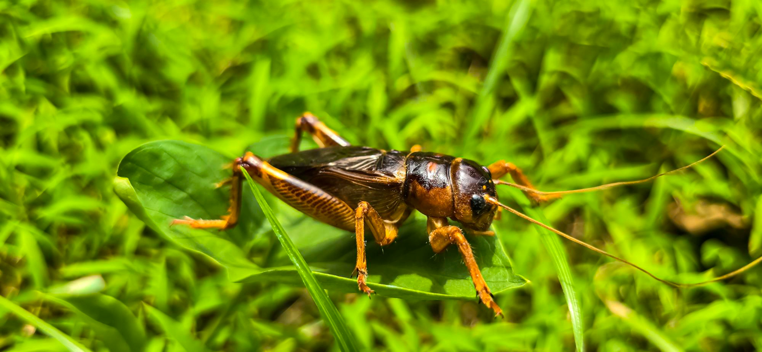
The Rise of Feeder Insects
Feeder insects like crickets, roaches, and worms have become a staple food for a wide range of exotic pets. As the demand for these insects increases, the feeder insect industry has grown into a multi-billion-dollar global business. However, the industry is vulnerable to disease outbreaks, which can not only impact insect populations but also disrupt the supply chain and affect the nutritional quality of the insects consumed by pet reptiles and amphibians.
Common Diseases Affecting Feeder Insects
Several pathogens including viruses, bacteria, fungi, and parasitic organisms threaten the health of crickets, roaches, and other feeder insects. Here’s a closer look at the most common species and their impact on insect populations:
Bacterial Infections
Bacillus thuringiensis
- Description: Bacillus thuringiensis is a bacterium commonly used as a natural pesticide. While effective in pest control, when present in high concentrations, it can infect feeder insects, leading to gastrointestinal issues and death (Roh et al., 2007).
- Hosts: Crickets, roaches, and other insects
- Symptoms: Gastrointestinal issues, weight loss, and death in overexposed insects.
Fungal Infections
Beauveria bassiana
- Description: Beauveria bassiana is an entomopathogenic fungus that infects insects by growing inside their bodies. Although used for pest control, it can cause severe damage when it infects high-density populations of feeder insects (Araújo et al., 2016).
- Hosts: Crickets, roaches, and many other insects
- Symptoms: Lethargy, body distortion, and death.
Metarhizium anisopliae
- Description: Metarhizium anisopliae is another fungus commonly used in biological pest control. However, in feeder insects, it infects the exoskeleton and internal organs, often leading to visible signs of body decay (Araújo et al., 2016).
- Hosts: Roaches, crickets, and others
- Symptoms: Body decay, loss of mobility, and death. The fungus often leaves orange-colored growths on the insect’s body.
Conidiobolus spp.
- Description: Conidiobolus species are fungal pathogens that can severely affect insect populations, particularly crickets and roaches. These fungi cause deformations and hinder the insects’ ability to function normally (Araújo et al., 2016).
- Hosts: Crickets and roaches
- Symptoms: Severe body deformation, loss of appendage functionality, and death.
Parasitic Eukaryotes
Gregarines (Protozoan Parasites)
- Description: Gregarines are protozoan parasites that infect the digestive systems of their insect hosts. They can significantly affect the insects’ growth and ability to feed, reducing their nutritional value (Gałęcki et al., 2019).
- Hosts: Crickets, roaches, and other insects
- Symptoms: Lethargy, reduced growth, and death. These parasites hinder the insect’s ability to consume food and grow properly.
Nematodes (e.g., Steinernema spp.)
- Description: Steinernema species are parasitic nematodes that infect insects, leading to paralysis and death. These parasites can also degrade the nutritional profile of the affected insect (Gałęcki et al., 2019).
- Hosts: Crickets and other insects
- Symptoms: Swelling, paralysis, and eventual death.
How Pathogens from Contaminated Insects Can Be Transmitted to Exotic Pets
Diseases affecting feeder insects are not just a threat to insect populations, they can also be transmitted to exotic pets. When reptiles and amphibians consume infected insects, they may become exposed to harmful pathogens, leading to illness or even death.
When a pet consumes an infected insect, the pathogen (such as bacteria, fungi, or parasites) can enter the pet’s digestive system. This can cause gastrointestinal issues or, in some cases, systemic infections that harm the pet. Protozoan parasites like Cryptosporidium can be ingested by pets and establish themselves in the pet’s gut (Mitchell, 2007). This can lead to internal infections that affect the pet’s health and growth.
The Importance of a Healthy Diet and Disease-Free Feeder Insects
Ensuring that the feeder insects provided to reptiles and amphibians are disease-free is essential to maintaining their health. This is why monitoring the health of feeder insects and practicing strict biosecurity measures in insect farming are vital to prevent the spread of harmful pathogens. By choosing trusted suppliers and ensuring that the insects are properly farmed and handled, pet owners can reduce the risk of disease transmission and ensure that their pets receive the safest, most nutritious diet possible.
Using Next-Generation Sequencing (NGS) to Monitor Feeder Insect Populations and Prevent Disease Spread
Next-generation sequencing (NGS) technology is revolutionizing how the feeder insect industry monitors the health of insect populations and detects pathogens before they cause widespread outbreaks. By sequencing the DNA of crickets and roaches, NGS can identify a range of viruses, bacteria, fungi, and parasites—even in the absence of visible symptoms. This early detection enables producers to take swift action, preventing significant damage to insect populations.
NGS allows for whole genome sequencing, identification of specific microbial species, and tracking genetic markers linked to disease resistance or susceptibility. Regular sequencing helps monitor microbial diversity, assess pathogen presence, and implement targeted interventions. Moreover, NGS data can reveal environmental factors contributing to pathogen outbreaks, enabling insect farmers to optimize their farming practices and minimize disease transmission. By adopting NGS, the feeder insect industry can enhance biosecurity, improve insect health, and create a more stable and sustainable supply chain.
Mitigating the Impact of Insect Disease on the Feeder Industry
The threat of disease in feeder insect populations is significant, impacting both insect health and the profitability of businesses that rely on them. Implementing strict biosecurity measures—such as maintaining clean, dry conditions, closely monitoring insect health, and using probiotics and natural remedies to manage diseases—can help mitigate pathogen spread. Additionally, understanding the specific needs of different insect species can improve farming conditions and reduce disease outbreaks.
Preventing disease outbreaks is essential not only for protecting industry profits but also for ensuring the safety and nutritional value of insects used to feed pet reptiles and amphibians. A healthy feeder insect population is crucial for the well-being of pets and the businesses that provide for them.
References
- Anankware, P. J., Fening, K. O., Osekre, E., & Obeng-Ofori, D. (2015). Insects as food and feed: A review. International Journal of Agricultural Research and Review, 3(1), 143-151.
- Araújo, J. P., & Hughes, D. P. (2016). Diversity of entomopathogenic fungi: which groups conquered the insect body?. Advances in genetics, 94, 1-39.
- Benson, K. G. (1999, April). Reptilian gastrointestinal diseases. In Seminars in Avian and Exotic Pet Medicine (Vol. 8, No. 2, pp. 90-97). WB Saunders.
- Gałęcki, R., & Sokół, R. (2019). A parasitological evaluation of edible insects and their role in the transmission of parasitic diseases to humans and animals. PloS one, 14(7), e0219303.
- Mitchell, M. A. (2007). Parasites of reptiles. Flynn’s parasites of laboratory animals, 177-216.
- Nyombe, R. P. M., Odhiambo, B., & Bulli, P. (2024). Bacterial and fungal microbes associated with cricket rearing systems in the tropics. Journal of Insects as Food and Feed, 1(aop), 1-14.
- Roh, J. Y., Choi, J. Y., Li, M. S., Jin, B. R., & Je, Y. H. (2007). Bacillus thuringiensis as a specific, safe, and effective tool for insect pest control. Journal of microbiology and biotechnology, 17(4), 547-559.
Categories: Bacterial Infections, Fungal Infections, Gastrointestinal Health, Next-Gen DNA Sequencing Technology, Pet Health, Safety and Wellness, Reptiles/Amphibians