Conjunctivitis, also known as an eye infection, commonly infects a wide variety of pet birds ranging from house finches to cockatiels, parakeets, and many more. Conjunctivitis can be caused by several different pathogens, which creates a highly variable disease presentation that makes conjunctivitis difficult to diagnose [1]. Considering the possibility of blindness and more severe outcomes, it is important for any bird owner to be aware of the signs of this difficult-to-diagnose disease.
Conjunctivitis infections are painful for your feathery friend and if left untreated can result in serious health complications and even death. If you suspect your bird has conjunctivitis, bring your pet to your exotic pet veterinarian as soon as possible.
What is Conjunctivitis?
Gram-positive bacteria such as Staphylococcus spp., Corynebacterium spp., and Clostridium botulinum, and Gram-negative bacteria such as Escheria coli, Chlamydia psittaci, and Mycoplasma spp., are causative agents of conjunctivitis [2,3]. Additionally, several fungi such as Aspergillus spp. and Candida albicans have also been associated with avian eye infections [3]. It is important to note that viruses, parasites, environmental exposures, trauma, foreign bodies, and Vitamin A deficiencies are also associated with conjunctivitis manifestation [4]. If a bird comes into direct contact with contaminated food or feces of an infected bird, conjunctivitis infections colonize conjunctiva, which is the membrane that surrounds the eye [5]. While conjunctivitis can affect all bird species, finches are particularly prone to infections. Symptoms of this disease include but are not limited to:
- Crusty eyes
- Swollen and red eyes
- Cloudy or glassy eyes
- Sinitis or upper respiratory infections
- Nasal and/or eye discharge
- Sneezing
- Deposits on cornea
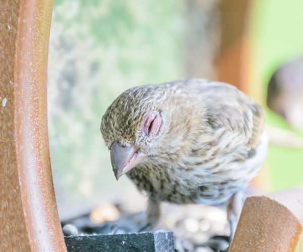
ild female house finch at a backyard feeder suffering from House Finch Eye Disease (also known as Mycoplasmal conjunctivitis).
Preventing Avian Conjunctivitis
Ideally, the best way to make sure your bird is void of a conjunctivitis infection is to ensure the bird’s breeder is reputable and upholds standard avian husbandry protocol. When a new bird is introduced to a household, the CDC recommends quarantining them for 30-45 days. Practicing preventative husbandry (proper cage positioning, daily cage cleaning, proper ventilation, etc.) is also critical in creating a low-stress environment so your bird is less susceptible to infection [6]. Regular veterinary visits are recommended as well.
Treating Avian Conjunctivitis
Your veterinarian will test your feathery friend to determine the strain of bacteria or fungus causing your pet’s infection. Saline flushes alongside topical and/or oral antibiotics are commonly used for treating conjunctivitis infections, and your veterinarian will determine the duration of treatment along with possible dietary supplements to aid in your bird’s recovery. Additionally, your veterinarian can help you identify possible lifestyle changes you and your bird can make to improve their quality of life and lessen the risk of recurrent infections. This entails understanding the exact pathogen that is impacting your bird, with modern technological advances allowing for more targeted clinical diagnostic interventions. In general, the prognosis for this infection is positive in mild to moderate disease presentations but is more variable in birds with severe symptoms.
Diagnosing Avian Conjunctivitis
Diagnosis of conjunctivitis infections is particularly complex, largely due to the overwhelming variety of causative pathogens involved, and consequently the difficulty culturing respective bacteria and/or fungi. While Gram-negative bacteria is already difficult to culture and often produces “no-growth” cultures, of equal concern is the rapid rate at which disease resistance in conjunctivitis-associated bacteria is increasing. For example, one study evaluating the antimicrobial resistance determinacy in Staphylococcus spp. recovered from birds increasing resistance to several different antibiotics dependent on the strain of the bacteria, underscoring the need to characterize the microbiome of avian eye infections [7]. Next-Gen Sequencing (NGS) technology is especially applicable for avian conjunctivitis due to the massive amount of variation in causative pathogens, highlighting the clinical applicability of using genomic sequencing to identify, analyze, and eventually treat birds more effectively.
The MiDOG All-in-One microbiome test may provide the answer to the diagnostic conundrum that conjunctivitis poses on your feathery friend. Utilizing NGS technology to detect and quantify all microbial DNA through untargeted and comprehensive sequencing and quantitative comparisons to reference databases, the MiDOG NGS technology provides a useful opportunity to shed light on the microbial makeup of your bird’s infection for clinical application. The MiDOG microbiome test is a microbial identification test grounded on scientific research that provides veterinarians DNA evidence for the guided treatment of bird infections, such as conjunctivitis.
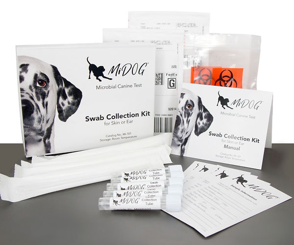
Find out if your vet uses MiDOG before you book your next appointment!
References
1. Altizer, S., Hochachka, W., & Dhondt, A. (2004). Seasonal dynamics of mycoplasmal conjunctivitis in eastern North American house finches. Journal Of Animal Ecology, 73(2), 309-322. doi: 10.1111/j.0021-8790.2004.00807.x
2. Ley, D. H., Hawley, D. M., Geary, S. J., & Dhondt, A. A. (2016). House Finch (Haemorhous mexicanus) Conjunctivitis, and Mycoplasma spp. Isolated from North American Wild Birds, 1994-2015. Journal of wildlife diseases, 52(3), 669–673. https://doi.org/10.7589/2015-09-244
3. Hawley, D. M., Moyers, S. C., Caceres, J., Youngbar, C., & Adelman, J. S. (2018). Characterization of unilateral conjunctival inoculation with Mycoplasma gallisepticum in house finches. Avian pathology : journal of the W.V.P.A, 47(5), 526–530. https://doi.org/10.1080/03079457.2018.1495312
4. Abrams, G., Paul-Murphy, J., & Murphy, C. (2002). Conjunctivitis in birds. Veterinary Clinics Of North America: Exotic Animal Practice, 5(2), 287-309. doi: 10.1016/s1094-9194(01)00002-0
5. Moyers, S., Adelman, J., Farine, D., Thomason, C., & Hawley, D. (2018). Feeder density enhances house finch disease transmission in experimental epidemics. Philosophical Transactions Of The Royal Society B: Biological Sciences, 373(1745), 20170090. doi: 10.1098/rstb.2017.0090
6. Amer, M., & Mekky, H. (2020). Avian gastric yeast (AGY) infection (macrorhabdiosis or megabacteriosis). BULGARIAN JOURNAL OF VETERINARY MEDICINE, 23(4), 397-410. doi: 10.15547/bjvm.2019-0035
7. Sousa, M., Silva, N., Igrejas, G., Silva, F., Sargo, R., & Alegria, N. et al. (2014). Antimicrobial resistance determinants in Staphylococcus spp. recovered from birds of prey in Portugal. Veterinary Microbiology, 171(3-4), 436-440. doi: 10.1016/j.vetmic.2014.02.034
Categories: Birds/Parrots, Exotic Pets, Eye Infections, Next-Gen DNA Sequencing Technology
