Next-Gen Sequencing can help diagnose bearded dragon upper respiratory infections.
Captive bearded dragons are particularly at risk for respiratory infections (like pneumonia), which are often caused due to a weakened immune system. Environment plays a key role in the pathogenesis of pneumonia, and so it is useful for pet owners to know the proper husbandry to take care of bearded dragons, which are the most popular pet reptile [1].
Upper respiratory infections are uncomfortable for your scaly friend and may result in death if left untreated. Early intervention is beneficial for treatment outcomes, and so if you suspect your pet has pneumonia, visit an exotic pet veterinarian immediately so they can diagnose and provide a treatment plan.
What causes Pneumonia in Bearded Dragons?
Although research into the respiratory microbiome of bearded dragons is limited, recent studies suggest the increased likelihood of concurrent viral and bacterial infections in persistent pneumonia cases [2]. Viruses associated with pneumococcal infections in bearded dragons include adenovirus, nidovirus, and more, while various bacteria such as Chlamydia pneumoniae and Mycoplasma spp. are also responsible [2,3,4]. Mycoplasma spp. are the smallest of all prokaryotic microbial cells, and do not have a cell wall [5]. The lack of a cell wall is particularly difficult for diagnostic purposes, because they do not stain with the Gram stain and occur in various distinct forms with a high variation in the size and shape of their cells and/or nuclei [2]. It is important for your veterinarian to identify the cause of your bearded dragon’s infection, because that will inform whether or not antibiotics are used.
For any reptile parent, identifying symptoms of pneumonia is critical. Symptoms include:
- Discharge from eyes or nose
- Bubbles from mouth or nose
- Rapid/shallow breathing
- Open-mouthed breathing
- Decreased appetite
- Lethargy
- Sneezing/snorting
- Weight loss due to decreased appetitive
- Puffing up their throat
In severe and prolonged cases, respiratory infections may advance to septicemia, which is a widespread infection of the bloodstream [6]. Consequently, it is important to visit a veterinarian as soon as you begin to notice these symptoms.

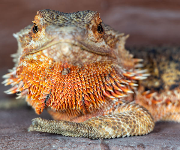
The image above depicts a healthy bearded dragon in a well-maintained terrarium.
Preventing Upper Respiratory Infections in Bearded Dragons
Pet reptiles are highly at risk for developing infections due to incorrect temperature and humidity levels. As the Manual of Exotic Pet Practice puts it, “the majority of diseases observed in captive reptiles are directly associated with improper husbandry” [7]. Notably, infections in captive reptiles are more common at lower ambient temperatures. In order to prevent upper respiratory tract infection in reptiles, it is important to reduce the viral and/or bacterial load while setting the proper temperature and humidity levels. A proper diet is also very important in maintaining optimal health for your reptile, as vitamin deficiencies can leave your reptile vulnerable to pathogens. And while it may seem obvious that cleaning your pet’s terrarium is important in warding off infections, cleanliness cannot be overstated. Isolating suspected cases of pneumonia from other reptiles is also necessary.
To learn more about proper husbandry habits for bearded dragons, make sure to visit this guide.
Treating Upper Respiratory Infections For Bearded Dragons
Considering the complicated nature of upper respiratory infections, it is important to consult with a veterinarian as soon as possible. Delay in care is painful and may result in septicemia and potential death. While treatment may entail antibiotics if there is a bacterial infection, in advanced cases, supportive therapy may be necessary in severe cases [6].
Your veterinarian will also help you identify possible lifestyle changes you and your reptile can make to improve their quality of life and lessen the risk of recurrent infections. This entails understanding the exact pathogen that is impacting your reptile, with modern technological advances allowing for more targeted clinical diagnostic interventions.
Diagnosing Pneumonia in Bearded Dragons
With the rise of antibiotic-resistant strains of pathogens, advances in recent clinical diagnostics have allowed for more targeted and efficient interventions. Historically, culture-based methods have been used to assess the nasal microbiome of reptiles, but there are notable diagnostic shortcomings. Specifically, Mycoplasma spp. are slow-growing and difficult to culture, producing numerous false negatives [2]. Recent studies have begun to shift away from problematic liquid cultures (that require multiple weeks of isolation) in favor of metagenomic sequencing.
Next-Gen Sequencing (NGS) has increasingly helped researchers and veterinarians characterize bearded dragon upper respiratory tract infections. A recent case study assessing pneumonia in a captive central bearded dragon with concurrent detection of helodermatid adenovirus and a novel Mycoplasma spp. used 16S ribosomal RNA gene sequencing with phylogenetic analysis to accomplish this [2]. This indicates the clinical applicability of using genomic sequencing to identify, analyze, and eventually treat reptiles more effectively.
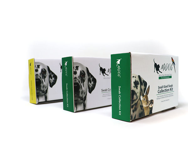
Despite its name, the MiDOG All-in-One Microbial Test may provide the answer to the diagnostic conundrum that upper respiratory infections pose for you and your reptile. Utilizing NGS technology to detect and quantify all microbial DNA through untargeted and comprehensive sequencing and quantitative comparisons to reference databases, the MiDOG NGS technology provides a useful opportunity to shed light on the microbial makeup of your reptile’s bacterial infection for clinical application. The MiDOG microbial test is grounded on scientific research that provides veterinarians DNA evidence for the guided treatment of reptile infections, such as pneumonia.

Find out if your vet uses MiDOG before you book your next appointment!
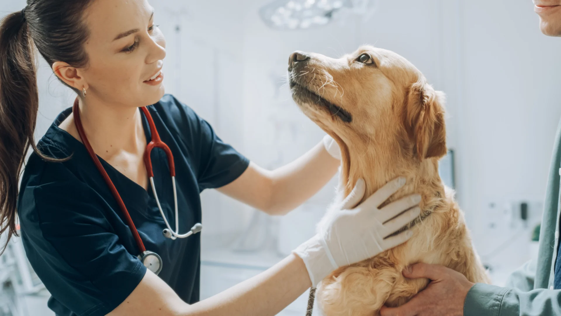
For health-related questions about your reptile or other exotic pet, reach out to a veterinarian that specializes in exotic pets.
References:
- Valdez, J., 2021. Using Google Trends to Determine Current, Past, and Future Trends in the Reptile Pet Trade. Animals, 11(3), p.676.
- Crossland, N., DiGeronimo, P., Sokolova, Y., Childress, A., Wellehan, J., Nevarez, J., & Paulsen, D. (2018). Pneumonia in a Captive Central Bearded Dragon With Concurrent Detection of Helodermatid Adenovirus 2 and a Novel Mycoplasma Species. Veterinary Pathology, 55(6), 900-904. doi: 10.1177/0300985818780451
- O’Dea, M., Jackson, B., Jackson, C., Xavier, P., & Warren, K. (2016). Discovery and Partial Genomic Characterisation of a Novel Nidovirus Associated with Respiratory Disease in Wild Shingleback Lizards (Tiliqua rugosa). PLOS ONE, 11(11), e0165209. doi: 10.1371/journal.pone.0165209
- Ebani, V. (2017). Domestic reptiles as source of zoonotic bacteria: A mini review. Asian Pacific Journal Of Tropical Medicine, 10(8), 723-728. doi: 10.1016/j.apjtm.2017.07.020
- Razin S. Mycoplasmas. In: Baron S, editor. Medical Microbiology. 4th edition. Galveston (TX): University of Texas Medical Branch at Galveston; 1996. Chapter 37. Available from: https://www.ncbi.nlm.nih.gov/books/NBK7637/
- Divers, S. (2020). Disorders and Diseases of Reptiles – All Other Pets – MSD Veterinary Manual. Retrieved 13 May 2022, from https://www.merckvetmanual.com/all-other-pets/reptiles/disorders-and-diseases-of-reptiles
- Deczm, M. M. D. M. P. & Tully Jr. DVM MS DABVP (Avian) DECZM (Avian), Thomas N. (2008). Manual of Exotic Pet Practice (1st ed.). Saunders.
Categories: Bearded Dragons, Exotic Pets, Reptiles/Amphibians, Respiratory Infection

