
Mycobacterium fortuitum is a rapidly growing nontuberculous mycobacterium (NTM) that is often overlooked as a pathogen. However, M. fortiutum has become increasingly recognized in veterinary and medical fields due to its association with a variety of infections in animals and humans. This organism, belonging to the same family as the causative agent of tuberculosis, shares some similar characteristics but differs significantly in terms of virulence and disease manifestation. While it is not the causative agent of classic tuberculosis, its ability to cause abscesses and other granulomatous diseases makes it a significant concern for pets.
Let’s explore the characteristics, clinical implications, and management of M. fortuitum infections for veterinary practice and its potential impact on pet owners!
What is Mycobacterium fortuitum?
Mycobacterium fortuitum is part of the Mycobacterium genus, which also includes M. chelonae and M. abscessus species. These organisms are characterized by their rapid growth rate, usually forming colonies within 7 days, and their ability to survive in a wide range of environmental conditions, including water, soil, and dust. Unlike the more well-known M. tuberculosis, M. fortuitum is not a primary pathogen but an opportunistic one, often affecting immunocompromised hosts or those with pre-existing conditions.
Mycobacterium fortuitum and Tuberculosis: Understanding the Difference
While Mycobacterium fortuitum and Mycobacterium tuberculosis both belong to the genus Mycobacterium, they differ significantly in their pathogenicity and clinical presentation. Tuberculosis (TB), caused by M. tuberculosis, is a major global health concern, characterized by its chronic and often severe respiratory disease. In contrast, M. fortuitum does not typically cause TB but is associated with localized infections such as abscesses, particularly in the skin and soft tissues.
Veterinarians should be aware that despite these differences, M. fortuitum can sometimes cause confusion in diagnosis due to the transient tuberculin skin sensitivity it may provoke. This reaction can occur in animals and humans exposed to the bacterium, leading to false-positive results in TB testing. Therefore, understanding the distinct clinical features and diagnostic approaches for these mycobacterial infections is crucial in veterinary practice.
A Link to Mycobacterium fortuitum in Fish?
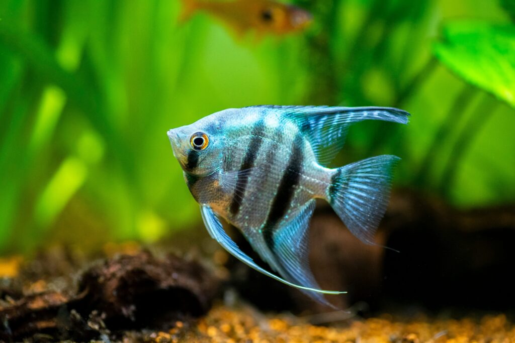
Mycobacterium fortuitum is well-documented in causing localized infections, particularly abscesses, in a variety of animals, including dogs, cats, and livestock. These infections often occur following trauma or surgical procedures, where the bacterium gains entry through wounds or compromised skin barriers. One of the most significant implications of M. fortuitum in veterinary medicine is its impact on aquatic species, particularly fish. Mycobacteriosis, caused by various species of Mycobacterium, including M. fortuitum, is a chronic disease that affects both wild and captive fish populations. The disease is characterized by the formation of granulomas, which can develop in various organs, leading to systemic infection.
In fish, the disease often presents as chronic wasting, with symptoms such as weight loss, lethargy, and skin ulcers. The presence of abscesses, particularly in the internal organs, is a hallmark of mycobacteriosis in fish. These abscesses are typically filled with caseous material and surrounded by a fibrous capsule, which can complicate treatment.
How to Identify Mycobacterium fortuitum
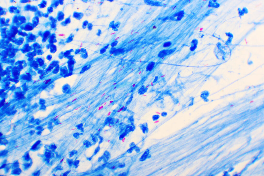
M. fortuitum infections typically present as non-healing abscesses or nodules, often accompanied by draining tracts. These abscesses are usually firm, with a caseous or purulent center, and may be mistaken for other bacterial or fungal infections. Cytology and histopathology of the lesions often reveal granulomatous inflammation, with acid-fast bacilli visible on special stains, confirming the presence of mycobacteria.
Diagnosing M. fortuitum infections requires a combination of clinical assessment, cytology or histopathology, and culture of the organism from affected tissues. Traditionally, polymerase chain reaction (PCR) assays targeting specific mycobacterial DNA can provide a diagnosis. However, one of the challenges veterinarians face is the potential for cross-reactivity between M. fortuitum and MAP in diagnostic tests, such as the tuberculin skin test or serological assays. Animals exposed to M. fortuitum may develop transient tuberculin skin sensitivity, leading to false-positive results when testing for Johne’s disease or bovine tuberculosis. Thus, Next-Generation Sequencing (NGS) such as the All-in-One Test offered by MiDog Animal Diagnostics, is a great way to more effectively identify not just Mycobacterium fortuitum, but all Mycobacterium species that may be present in a fish!
How to Manage and Treat Mycobacterium fortuitum Infection in Pet Fish
For veterinarians working with aquaculture or pet fish, managing mycobacteriosis can be challenging due to the bacterium’s resilience and the difficulty in treating affected fish. The primary approach to controlling the spread of M. fortuitum in fish populations involves:
- Strict quarantine of new fish
- Regular water quality monitoring, and
- Prompt removal of infected individuals.
Treatment options are limited, as Mycobacterium spp. are generally resistant to many common antibiotics. Long-term administration of antimicrobials such as clarithromycin, rifampin, and ethambutol has been used with varying degrees of success, but complete eradication of the infection is rarely achieved. Consequently, prevention remains the most effective strategy in managing mycobacteriosis in fish. Additionally, surgical excision of the abscesses, combined with prolonged antibiotic therapy based on culture and sensitivity results, is often necessary. However, due to the chronic nature of these infections, long-term follow-up and management are essential to prevent recurrence.
Conclusion
Mycobacterium fortuitum, though not as notorious as Mycobacterium tuberculosis or Mycobacterium avium subspecies paratuberculosis, plays a significant role in veterinary medicine due to its ability to cause a range of infections across different species. From its impact on fish populations to its role in abscess formation and its potential to cause diagnostic confusion with other mycobacterial diseases, understanding this opportunistic pathogen is crucial for veterinarians.
For pet owners, particularly those with aquatic pets or animals prone to infections, awareness of Mycobacterium fortuitum and its potential risks is essential. Early recognition and appropriate management of infections can prevent long-term complications and improve outcomes for affected animals.
Veterinarians are encouraged to stay informed about the latest developments in mycobacterial research and to employ a comprehensive diagnostic approach when dealing with suspected mycobacterial infections. Through diligent care, accurate diagnosis, and effective management, the challenges posed by Mycobacterium fortuitum can be successfully navigated, ensuring the health and well-being of both animals and their owners.
References
Falkinham, J. O. (2016). Environmental sources of nontuberculous mycobacteria. Clinics in Chest Medicine, 36(1), 35-41.
Gauthier, D. T., & Rhodes, M. W. (2009). Mycobacteriosis in fishes: a review. The Veterinary Journal, 180(1), 33-47.
Tortoli, E. (2014). Microbiological features and clinical relevance of new species of the genus Mycobacterium. Clinical Microbiology Reviews, 27(4), 727-752.
Runyon, E. H. (1959). Anonymous mycobacteria in pulmonary disease. Medical Clinics of North America, 43(1), 273-290.
Categories: Exotic Pets, Fish, Fungal Infections, Marine Mammal, Next-Gen DNA Sequencing Technology